The Final Question: To Drink, or Not to Drink?

As mentioned in the last blog post, the impacts of alcohol on the growing fetus can be controlled by the genes. It is widely known that the symptoms of all the disorders that fall under the title Fetal Alcohol Spectrum Disorder fall on a gradient. However, there is not a lot of grey area in regards to prenatal alcohol exposure; the mother either drinks or she doesn’t. A child can’t be sort of exposed to alcohol in utero, the same way a woman can’t be sort of pregnant. The question then is, why is there a gradient when of symptoms when the actual exposure is so definitive? A study done by Hemingway et al. looks at the effects of fetal genetics in cases of FASD in an attempt to answer this question. If two fetuses were exposed to the same level of alcohol at the same time during pregnancy, but showed vastly different symptoms, it would have important implications for public health and education surrounding maternal drinking. The purpose of this study was to compare ranges of FASD symptoms among four groups; identical twins, fraternal twins, full siblings and half siblings sharing the same birth mother. The study was attempting to do the following four things;

  1. Determine if the prevalence of FASD was higher among fraternal twins compared to identical.
  2. To determine if the prevalence of FASD increases with the decrease in shared genetic material between sibling pairs.
  3. Document the greatest difference between twin pairs with identical Prenatal Alcohol Exposure (PAE). Can twins with identical PAE be at the opposite ends of the FASD spectrum?
  4. Estimate the heritability of the FASD phenotypes (the physical presentation of the symptoms)

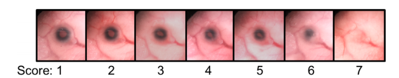
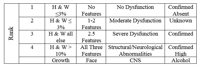
The presence of FASD among the sibling pairs was evaluated based on the following criteria (in the presented order);

  1. Growth deficiency
  2. FAS facial phenotype (thin upper lip, wide set eyes, etc)
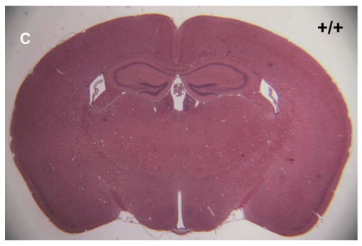
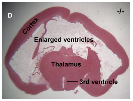
  3. CNS structural or functional abnormalities (behavioural disorders, misculoskeletal issues, etc)
  4. PAE

The degree of each feature was ranked on a four-point scale. 1 is the absence of the criteria, and 4 is a strong, “classic” presentation of the FAS phenotype.

The four-point scale used to grade the FAS criteria. The scores are reported as the numbers, 4334 for example. 4334 would be a 4 in the growth category, a 3 in the facial category, a 3 in the CNS category and a 4 in the alcohol category.


The siblings were placed into four groups;

  1. Monozygotic twins (identical twins, share 100% of their genome)
  2. Dizygotic twins (fraternal twins, share ~50% of their genome)
  3. Full- siblings (share ~50% of their genome)
  4. Half-siblings sharing a common mother (share ~25% of their genome)

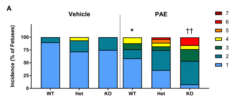
The results showed the proportion of sibling pairs with FAS was very similar to the proportion of their genome they shared; all the monozygotic twin pairs shared FASD, 56.4% of the dizygotic twin pairs shared FASD, 40.7% of full siblings and 22.2% among half siblings. This represents a linear decrease with the proportion of genome shared. There was no information that suggested gender impacted the diagnosis of FASD.

Although the diagnosis of FASD was present in the approximate proportion as the shared genome, it did not always present itself to the same degree. The concordance (similarity) between the dizygotic twins was extremely varied. One twin could experience low to moderate symptoms of FASD, while the other could experience severe symptoms as reported by the four-point scale. The full-siblings pairs and half-sibling pairs also experienced large ranges of the FASD phenotypes.

Despite the difference in the FASD presentation, both monozygotic and dizygotic twin pairs had a 100% heritability rate. This means that the presence of FASD was entirely due to their genome (despite the differences in phenotype) and not due to environmental risk factors.

The results of this study support the idea that fetal genetics can impact the impacts of prenatal alcohol exposure. The prevalence of FASD among the sibling pairs was directly correlated with how much of their genome was shared between them. Similar studies done over the years, both on twins and on model animals, with FASD has also presented with similar results. This suggests that there is a genetic component to FASD. Although the genes may influence the presentation of the symptoms, they can not cause them.

It gives further support to the fact that FASD in all its forms is 100% preventable. The answer to the question; What is a safe amount of alcohol to drink while pregnant? Has a very simple answer: nothing at all.

Article Reference

Hemingway, S.J.A., J.M. Bledsoe, J.K. Davies, A. Brooks, T. Jirikowic, E.M. Olson & J.C. Thorne (2019). Twin study confirms virtually identical prenatal alcohol exposures can lead to markedly different fetal alcohol spectrum disorder outcomes- fetal genetics influences fetal vulnerability. Advances in Pediatric Research. 5(23).

Is FAS Primarily Under Genetic Control?

If a mother consumes alcohol during her pregnancy, she puts her unborn child at risk of facial anomalies, neurodevelopmental delay and even spontaneous abortion. Although the prevalence of FAS in the US is approximately 0.2-2.0 cases per 1,000 live births, FAS actually occurs in less than 10% of pregnancies, even if the woman drinks heavily throughout. If FAS does not impact all pregnant woman equally or to the same degree, then understanding why some women have increased risk factors for FAS is crucial. There are many cellular events during development that can be impacted by the presence of alcohol, which includes cell death, decrease in cell binding and impacts to molecules that induce growth in the embryo. It seems that the malformations that impact the fetus shows organ selectivity.

The damage induced by prenatal alcohol (PAE) is not hereditary. That means it can not be passed down from the mother to her child, the damage arises from direct contact with toxic substances during development. However, there does seem to be a genetic component to the severity of the damage that occurs. This may be why two mothers can drink the same amount at the same time during pregnancy, and one child will have severe effects and one child will have little to no impact. In a study done on mice, the severity of the effects was correlated with the alcohol concentration in the dam’s blood. The alcohol concentration would depend on the levels of alcohol dehydrogenase[ADH (an enzyme that assists in the breakdown of alcohol)], which would be controlled at the genomic level (by the genes). This suggests that mother’s who metabolize (break down) alcohol at different rates may lead to differences in fetal alcohol exposure, and differences in the level of resulting FAS symptoms.

The alcohol metabolism pathway. Ethanol (alcohol) is ingested, and the alcohol dehydrogenase removes a hydrogen from the alcohol to convert it to Acetaldehyde. The Acetaldehyde dehydrogenase (a second enzyme) converts Acetaldehyde into Acetic acid, which is vinegar. From Acetic acid, it is converted into carbon dioxide and water, and removed from the body.

After a mother drinks alcohol, it is quickly absorbed by the intestines. The rate of absorption depends on the amount, the concentration and the timing of the drink. Over 90% of the ingested alcohol is metabolized by the liver. The genes that code for the enzymes that remove the alcohol in the liver vary in their catalytic activity. This means the rate at which the enzyme works to remove the alcohol can vary depending on the genes that control it. These features and their different frequency in various ethnic populations may act as a protective/enhancing factor towards alcohol abuse and alcohol-related pathologies, such as FAS.

A study done on a mixed race population in South Africa found a prevalence of 39-46 FAS cases out of 1,000 births. They found a variation on the allele that codes for ALDH (ALDH2*2). It seemed that either a mother or a fetus with the ALDH2*2 variation had a protective quality against FAS, and those with the other variations on the gene did not have the same protective factors. It is important to note that the ALDH2*2 is more catalytically active than the other variants. This means the enzyme coded for by the ALDH2*2 works to break down alcohol faster than the other variations of the gene. Breaking down the alcohol consumed by the mother faster, would decrease the concentration of alcohol the fetus is exposed to, and possible provide protective factors against the effects of FAS. When the mothers took a breathalyzer test, it was found the FAS mothers had a higher breath alcohol concentration compared to the non-FAS mothers.

An example of the placental barrier and how substances from the mother’s blood can be passed into the fetus.

When
a pregnant woman ingests alcohol, it is delivered directly to her fetus through the placental barrier. The placental barrier is not a natural barrier to alcohol, and has the ability to let alcohol pass into the fetal circulation. Although the placenta does have enzymatic activity, the levels are much lower in comparison to the levels present in the mother’s blood. The fetus can remove some toxins (like alcohol) from the blood on their own, but not on the same scale or at the same rate as the mother could. By having a decrease in the enzymatic activity, the fetus would take longer to remove the alcohol from their blood, and the longer the alcohol in present in their blood stream, the greater the possibility of ending up with alcohol-related birth defects.

There are many potential factors that go into a child developing FAS. However, by examining the genetic basis behind it, more education and awareness can be generated about potential causes of the disease. More research would be needed to determine if the genes impacted are controlled by multiple factors, or just one.

Article Reference

Gemma, S., S. Vichi & E. Testai (2007). Metabolic and Genetic Factors Contributing to Alcohol Induced Effects and Fetal Alcohol Syndrome. Neuroscience and Behavioural Reviews. 31(2): 221-229.

Mice and Genes and Alcohol, Oh My!

Using animal models is an incredibly effective way of discovering the function of particular genes. Studies can be done on animals that can not be done on humans for ethical reasons. This study, conducted by Boschen et al. looks at the function of the meiosis-specific nuclear structural protein 1 (MNS1). The function of this protein is relatively unknown, and this study aims to look at its function in the facial, eye, brain and spinal cord defects that are common in humans with prenatal alcohol exposure (PAE). This is done in mice in a way that mimics first trimester alcohol exposure. Studies on the MNS1 protein have mainly focused on sperm, but it has been noted that knockout mice (mice who have had the gene removed from their DNA) have a higher rate of characteristics typically associated with PAE. MNS1 plays a role in motile cilia, which help in the circulation of signalling molecules that help form the axis of a body. The authors hypothesized that alterations to MNS1 could contribute to the development of craniofacial dysmorphologies, such as those characteristic of fetal alcohol syndrome (FAS). In lay terms; the authors believe that if you alter the MNS1 protein, it will cause facial deformities in the mice that are similar to the deformities seen in humans with FAS.

The subject mice were given two injections of ethanol (consumable alcohol) at a time in pregnancy that mimics the first trimester in humans. The maternal blood alcohol peaked at an average of ~440mg/dL. The control group was administered a similar concentration of a benign solution. After the injection, the dams were allowed to gestate normally until GD17. The dams and fetuses were then euthanized.

The eye deficits were analyzed and graded on a scale from 1 to 7, which can be seen below.

The gradient of eye deformities that were found in the fetuses. 1 represents a normal eye, 2-7 represent abnormal eyes.
A typical brain can be seen on the left, a case of hydrocephalus can be seen on the right. In the case with hydrocephalus, the excess cerebrospinal fluid can be seen expanding into the brain.

Brain tissue was removed from the fetuses and assessed for hydrocephalus. Hydrocephalus is a build up of cerebrospinal fluid in the brain. Normally, this is stored in special areas called the ventricles and circulated, but in a case of hydrocephalus, it accumulates and puts pressure on the brain.

Overall, it was found that fetuses with a knockout of the MNS1 gene that codes for the protein, or a defect to the MNS1 gene were more prone to facial defects, eye defects and hydrocephalus compared to the controls. There were no significant differences between the PAE group and the controls in terms of length or weight.

This is a sample of a normal brain from the mice at GD17. You can see the three ventricles (where the cerebrospinal fluid is stored) are normally sized, and the cortex (the pink area) has all the correct structures in the correct space. The cortex is responsible for controlling brain function and communication.

In this image, you can very see the expansion of the ventricles and the excess cerebrospinal fluid. The light space in the middle are the ventricles, which have merged together. You can also see an extreme reduction in the cortex, with little midbrain structures remaining.

A comparison of eye defects in the control group (vehicle) and the group injected with ethanol (PAE).

The above graph was generated by the authors and included in the paper. WT stands for wild type, which is a mouse that has no alterations made to the gene that codes for the MNS1 protein. Het stands for heterozygous. There are typically two copies of a gene that will code for a protein. With the heterozygous mice, they had one copy of the gene coding for the MNS1 protein altered, and one that was left completely normal. KO stands for knockout, these mice have had the gene coding for the MNS1 protein completely removed. The coloured scale on the right is the gradient of eye deformities, which is the same scale as the one showed before. As you can see in the graph, the mice with PAE have higher instances of more severe eye deficits compared to the control group.

In mice with eye deficits, there was also an increase in craniofacial deformities that can be correlated with the facial deformities seen in humans with FAS.

The results of this study suggest that the loss of the Mns1 gene increases the susceptibility of eye, craniofacial and central nervous system deformities.

The deformities may be caused in part by the disruption of proper motile cilia function. In normal development, cilia distribute morphogens (a substance that helps determine the pattern of tissue formation) to develop the left and right axes. In previous studies, mice with knock out MNS1 proteins have increased cases of situs inversus. Situs inversus is a condition where organs can develop in a mirrored position to what is typical.

The diagram shows a normal person on the left and a person with situs inversus on the right. The liver can be used as a landmark; in a normal patient it is on the left (remember the diagram is showing a person facing you, ie their right is your left) and in a person with situs inversus the liver is on the left.

This study is important because it is the first study done looking at the Mns1 gene and its association with the symptoms of FAS. Although it is done in a model organism, the results can potentially be applied to humans. Knowing this, new therapeutic technologies can be created and implemented that directly targets this pathway. Also, preventative measures can be taken in utero if a mother is drinking during pregnancy that may help to correctly form the motile cilia.

Article Reference

Boschen, K.E., H. Gong, L.B. Murdaugh & S.E. Parnell (2018). Knockdown of Mns1 Increases Susceptibility to Cranfiofacial-Defects Following Gastrulation Stage Alcohol Exposure in Mice. Alcoholism; Clinical and Experimental Research. 42(11):2136-2143.

Do Patients with FASD Suffer in Silence?

Fetal Alcohol Spectrum Disorders (FASD) can encompass a wide range of physical, cognitive and behavioural deficits due to the prenatal alcohol exposure (PAE). One area currently under heavy investigation is neurobehavioural function, specifically a patient’s communication abilities. Neurobehavioural function is how the nervous system (ie. the brain) relates to a person’s behaviour. Studies, for example the one done by Doyle et al., are looking at both functional and social communication, which is one’s ability to exchange information and interact with others. Young children with FASD experience communication impairment, which can drastically impact quality of life. Previous studies have found that communication skills in children and adolescents worsen with age, which suggest that communication skills are arrested in development, rather than just delayed.

Communication abilities encompass a wide range of social and personal skills.

Individuals with FASD struggle to meet the communication demands required by society, which requires intact social cognition, executive functioning, and language skills. This study examines the relationship between cognitive variables and a patients communication abilities. By exploring the communication ability in patients with PAE, it will help determine the cognitive mechanisms that contribute to communication deficits, which could lead to improvements in therapies and preventative measures.

The authors explored whether or not cognitive variables could predict the communication ability among adolescents with heavy PAE. They hypothesized that:

  1. Cognitive variables (such as working memory and language) that are shown to be predictive in other neurodevelopmental disorders would significantly predict communication ability in alcohol exposed youth and;
  2. These cognitive variables would show differential relationships with communication ability between alcohol exposed youth and typically developing controls.

This study was done as part of the Collaborative Initiative on Fetal Alcohol Spectrum Disorders, Phase Three (CIFASD III), which is a comprehensive battery of studies on neuropsychological and behvaioural measures in adolescents with FASD. Adolescents in the study were between the ages of 10 and 16 years old, and were separated into a PAE group (n= 1420 and a control group (n= 160). Control subjects were recruited from the same communities as the PAE groups. Measures assessed the relation between communication and performance in three areas; working memory, executive function and language.

Working memory involves storing, focusing and man

Multiple tests were administered that assessed socialization, daily living skills, spatial working memory and executive function. The tests were a mix of caregiver reports and neuropsychological assessments with the adolescent.

The results of the tests showed that the adolescents in the alcohol exposed group had a lower general cognitive ability, lower communication ability, and lower working memory compared to the control group. The alcohol exposed group showed a significantly lowered rate of cognitive and communication ability compared to the control group overall.

A graph generated by the authors showing the interaction between communication ability and word generation score in alcohol exposed and control adolescents.

The area where there was the most significant difference was in the Word Generation score. Here, participants were required to “…name as many words that fall into a certain category… or start with a certain letter in 60 seconds” to assess how their behaviour controlled their language.

A classic verbal reasoning question. One must combine the process of the answer given with the different problem in order to find the correct answer.

Overall, these results suggest that there is a difference in the communication abilities of adolescents exposed to alcohol in utero in comparison to a typically developing control group. The verbal reasoning aspect of the developmental delay is shown to impact the PAE group more strongly. Verbal reasoning is the ability to combine multiple pieces of information to produce one coherent piece of speech. Essentially, this means that patients with FAS have difficulty retrieving the words. They know what they want to say, but they can’t find the word in their mind in order to be able to produce a coherent thought or sentence.

The results of this study could be used to target speech and language therapies to have a greater impact on the aspects of language development most impacted by the alcohol exposure. Although the process of language comprehension and speech production is complex, any insight can help to improve the quality of life of someone experiencing the symptoms of Fetal Alcohol Syndrome.

Clinical interventions may also help to improve the communication deficits. Previous studies have shown that therapies directed at verbal reasoning have also shown improvements in self-regulation, attention and certain academic skills.

Article Reference

Doyle, L.R., E.M. Moore, C.D. Coles, J.A. Kable, E.R. Sowell, J.R. Wozniak, K.L. Jones, E. P. Riley & S.N. Mattson (2018). Executive Functioning Correlates with Communication Ability in Youth with Histories of Heavy Prenatal Alcohol Exposure. Journal of the International Neuropsychological Society. Doi: 10.1017/S1355617718000772

The answer to the verbal reasoning question: JR is to IP as NJ is to MH (a -1-2 rule)

Impacts of FAS Post-Adolescence

It is well known that there are certain psychological and social impacts as a result of in utero exposure to alcohol. Some of the symptoms include reduced cognitive functions, impaired verbal comprehension and certain behavioural issues, which may include rule breaking and aggressive behaviour. The results of these symptoms means that adolescents may have trouble in school or even issues with the law.

Despite all of the information present on the impacts of alcohol during childhood and throughout adolescence, there have been few studies done on how these symptoms impact someone with FAS during adulthood. A novel study done by Rangmar et al. predicted that the FAS study group would experience more problems in school, mental illness, and alcohol and illicit drug abuse.

All the participants in this group were required to have all four of the common features of FAS; history of alcohol abuse while in utero, characteristic pattern of facial abnormalities, growth deficits and abnormalities of the central nervous system. The participants had a mean age of 32 years. The subject group was 37% female and 63% male. It is important to note that 81% of the adults in the FAS group had been placed in state care as children.

Overall, it was noted that participants in the FAS group had a lower level of education, they were less likely to have a biological child, had higher rates of crime, and were more likely to commit a severe crime.

A comparison of the FAS study group with the controls.

The FAS group also had a higher rate of treatment for an alcohol-related disorder, drug abuse, treatment for a psychiatric disorder, self-inflicted injury and received at least one psychotropic drug.

The results of this study show that adults with an FAS diagnosis have a higher level of secondary disabilities, which supports the authors overall hypothesis. The FAS adults experienced more trouble in school, limited career options, and had higher rates of drug and alcohol abuse.

This study was important in showing the results of FAS beyond adolescents. There are so many studies focusing on the impacts on children, and it is often forgotten that FAS is a lifelong disease. From the results of this study, one can infer that the symptoms of FAS do not disappear over the course of a lifetime, instead they actually seem to be exacerbated by the independence given in adulthood.

This study is limited in the fact that the subjects and controls are taken from a single country, so the data can not be extrapolated to a worldwide scale. A future study should try and take a broader perspective, to see if the data acquired from this study can be applied to patients with FAS worldwide.

Article Reference

Rangmar, J., A. Hjern, B. Vinnerjung, K. Stromland, M. Aronson & C. Fahlke (2015). Psychosocial Outcomes of Fetal Alcohol Syndrome in Adulthood. Pediatrics. Doi: 10.1542/peds.2014-1915

The Darker Side of FASD

In the United States, suicide is one of the leading causes of death for adolescents between the ages of 13 and 18. Patients with symptoms falling on the FASD spectrum make up approximately 5% of youth in the United States, and as a result, they too experience suicidal thoughts and ideations. This is exacerbated by the fact that 94% of adolescents and adults with pre-natal alcohol exposure (PAE) have some form of a mental health problem. These problems are underscored by the degree of suicide risk in adulthood, with 43% of patients reporting suicide ideation and 23% reporting a history of suicide attempts.

Although many studies have been done on FASD adolescents with a lower IQ, and how that relates to their risk of suicide, nothing of the sort has been done for higher functioning FASD patients. This study, done by O’Connor et al., examined the prevalence of suicidal ideations and serious suicide attempts in adolescents with high functioning FASD.

This study involved 54 adolescents between 13 and 18, with 15.69 being the mean age in years. The mean IQ was 91.11. Each participant was tested for classic FASD symptoms and criteria, which includes growth retardation, FAS facial deformities (such as a flat lip and wide set eyes), neurodevelopmental dysfunction and exposure to alcohol in utero. The three main classifications of FASD found in the participants were Fetal Alcohol Spectrum Disorder, partial Fetal Alcohol Spectrum Disorder and Alcohol Related Neurodevelopmental DIsorder.

The suicide risk for the participants was recorded using the Children’s Interview for Psychiatric Syndromes (ChIPS). This was used to identify adolescents who experienced suicidal ideations and or who had made a previous suicide attempt. For this study, ideation was classified as a participant having any thoughts about killing themselves or verbalizing a suicidal intention. A suicide attempt was classed as an instance of deliberate harmful behaviour with a conscious or clear intent of a wish to die, with medical attention following an attempt.

The results of this study revealed that 35.2% of the adolescents had suicidal ideations and 13.0% had made at least one suicide attempt in the past twelve months. There was no significant difference between males and females who expressed ideations of suicide, but there was a significant difference in the suicide attempts between the sexes. 29.2% of males attempted suicide while 0% of females had a suicide attempt. Analyzing the IQ’s, it was found that male’s who attempted suicide had a significantly lower IQ compared to non-attempters. 36.8% of adolescents who reported suicide ideation also met the criteria for depressive disorders. Among the male sample, 42.9% reported a serious suicide attempt.

Comparing the results of this study to the national average, it is found that adolescents with FASD have a 5.5 times greater chance of making a serious suicide attempt compared to other adolescents of their age range. The results of the sex difference was even more drastic; nationally, typically-developing males have a suicide attempt rate of 1.5%. The rate of suicide attempts for males with FASD was 19.5 times higher at 29.2%.

The results of this study highlight a need for more monitoring and interventions for individuals with FASD. It gives support for the fact that FASD is not just a developmental disorder, there are serious social and behavioural issues associated, which can lead to extreme consequences for the adolescents impacted. It also poses the question of why females are less impacted, and what protective factors do they possess that males do not?

Teens and adolescents with FASD are at a much greater risk of developing life-threatening ideations compared to the rest of the population, with males being a significantly higher risk. The results of this study should provide motivation to implement some type of suicide screening for adolescents in general, and not just adolescents with FASD.

Article Reference

O’Connor, M.J., L.C. Portnoff, M. Lebsack-Coleman & K.M. Dipple (2019). Suicide risk in adolescents with fetal alcohol spectrum disorders. Birth Defects Research. Doi: https://doi.org/10.1002/bdr2.1465

On Epigenetics…..

Epigenetics is the study of heritable phenotypic changes that do not involve alterations to the DNA sequence. In English: epigenetics mainly deals with factors that have the ability to turn genes on or off.

Epigenetics can be influenced by many factors outside of the genes.

Any cell that is exposed to a toxin will likely result in harmful effects for the cell. However, the cells that are at the most risk are the ones that are still in the developmental process. This means that when a developing embryo or fetus is exposed to alcohol, it is highly impacted because it’s entire body (and therefore the entirety of it’s cells) are developing while being exposed to the alcohol. The cells can become damaged, and then as the embryo or fetus grows, the damaged cells will produce lineages of cells are also impacted by the alcohol. This is why an embryo in an earlier stage of development will be impacted to a greater degree in comparison to an embryo or fetus at a later stage.

Although FAS can be linked to genetic factors and maternal metabolism, there is strong evidence for epigenetic factors as well.

Two of the main players in epigenetics are metabolites and metabolic enzymes. Metabolites are the intermediate products of a reaction that breaks down an initial product into something that can be eliminated from the body. Enzymes are biological molecules which function to speed up a reaction, so metabolic enzymes will increase the rate at which a substance is broken down into metabolites. If more of an enzyme is available, then the substance, alcohol for example, will be broken down faster. Why does this matter? Well, the faster the alcohol can be broken down in the mother’s system, the less chance it will have of reaching the embryo’s system and causing alcohol related damage.

Epigenetic mechanisms have the most influence over an embryo in the early stages of pregnancy. Many studies have shown that alcohol is particularly prone to targeting protein modifications, particularly histones.

The hierarchy of genetic material organization.

Histones are the major structural protein of the chromosomes. The DNA is wrapped around the histones in order to condense it so it may take up less space in the cell. There are three main ways the hihstones can be modified; acetylation, methylation and phosphorylation. Alcohol has the ability to affect the histones in all three modified forms. Since the replication of DNA, as well as the proteins DNA codes for, is so essential during the developmental stages, the impacts alcohol can have on the histones can be extremely detrimental.

The acetylation of a histone occurs when an acetyl group is added to one end of the histone protein. The addition of this acetyl group causes the DNA wrapped around the histone to become more “relaxed”. The relaxed DNA is easier to transcribe and results in an increased rate of gene transcription. Gene transcription can involve anything from making more proteins essential for development, or turning other genes on and off in order to facilitate other developmental processes.

When the histones are acetylated, as seen on the bottom, they open up and have more of the DNA exposed instead of tightly wrapped around them .

In utero alcohol exposure can cause hyperacetylation or hypoacetylation of the histone proteins. Hyperacetylation increases the amount of acetyl groups added to the histones. With more acetyl groups added, more of the DNA wrapped around the histones will enter into that “relaxed” state, and allow for more gene transcription. In previous studies done, the hypoacetylation impacted genes are the genes that are responsible for fetal heart development.

Histone modifications can also occur via methylation and phosphorylation. These involve the addition of a methyl group or a phosphate group to the histone proteins, and can have affects similar to the acetylation.

Although not much is known about how epigenetics are impacted by alcohol exposure, it has been supported that alcohol exposure can impact the histone modifications and lead to an over or under-activation of certain genes or signalling pathways. However, it is not known how these genes play a role in the symptoms seen in patients with FASD.

Despite this not being a concrete source of information on the causes of FASD symptoms, it still gives insight into the dangers of in utero alcohol exposure.

Article Reference

Chanchal, M., D. Halder, K.H. Jung & Y.G. Chai (2017). In Utero Alcohol Exposure and the Alteration of Histone Marks in the Developing Fetus: an Epigenetic Phenomenon of Maternal Drinking. The International Journal of Biological Sciences. Doi: http://www.ijbs.com/v13p1100.htm

Is FAS in children with PAE linked to ADHD?

This study by Pagnin et al. investigated if there was a relationship between alcohol consumption during pregnancy and mental disorders in children. The overall prevalence of women who consume alcohol has increased, and as a result, so has the number of women who consume alcohol while pregnant. One in ten pregnant women consume alcohol, and one in five that do, drink alcohol at a level that poses a serious risk to their unborn child. Although at the extreme end of the fetal alcohol syndrome spectrum are deformities of the face and head and dysfunction of the central nervous system, more subtle consequences can arise that fall under the category of alcohol-related neurodevelopmental disorders (ARND). Children with ARND are characterized as having behavioural or cognitive abnormalities. Cognitive abnormalities are mental disorders that typically impact learning, memory and problem solving.

Even though one normally wound’t think of PAE impacting the genes, it impacts the developmental processes of the child, which can impact the genetic code to a degree.

The link between low alcohol consumption and mental disorders is not as clear as with a high alcohol consumption. The interactions with their environment after a child has been born can be just as effective at causing a mental disorder as prenatal alcohol exposure (PAE). The link then between low to moderate alcohol consumption and mental disorders can be thought of as being part of a broad nature vs. nurture debate. Is it solely the alcohol that caused the disorder? Is it solely the postnatal environmental factors? Or is it a combination of both?

The purpose of this study was to investigate the patterns of alcohol consumption in twelve-year old children, specifically focusing on episodic and regular use of alcohol as risk factors for a child developing ADHD.

This study was longitudinal and prospective, which means it was conducted in a way that involved repeated observations of the study population over a period of time. The study started with 449 pregnant women attending a specific clinic and had no medical complications. After twelve years, only 81 mother-child pairs attended the scheduled psychiatric interview. The presence of mental disorders was assessed in the children by giving them the Kiddie Schedule for Affective Disorders and Schizophrenia for School-Aged Children. This is a large collection of symptom rating scales and diagnostic criteria for psychiatric conditions. It is presented as a set of screening questions and the given answers are used to determine if a mental disorder is present, and if it is, the severity of the present symptoms.

Mothers were also given tests; a Self-Reporting Questionnaire to identify any non-psychiatric disorder and the Alcohol Use Disorders Identification Test, which determines if there is any alcohol problem, both past and present. Other tests were also administered to determine if there was any risk drinking during pregnancy, if there is any dependence, and the number of trimesters alcohol was consumed.

Statistical analyses were done to determine the relationships between all the variables and to see if there was a connection between PAE and the presence of ADHD.

Of the children who were sampled with the questionnaire, 45.6% were identified as having some kind of mental disorder, with 22.8% of mental disorders being externalizing and 32.1% being internalizing. A child with an internalizing disorder will keep the problems to themselves, and a child with an externalizing disorder will have symptoms or behaviours that are visible to an observer. ADHD made up 25.9% of the mental disorders diagnosed in the all the children.

Out of 81 children, a total of 21 had ADHD. ADHD was present in 13 of the 35 males tested, and in 8 of the 46 females tested.

ADHD was considered an externalizing disorder, and of the 26 children with externalizing disorders, 81% had ADHD. Compared to children without ADHD, children with ADHD had a significantly greater proportion of mother’s who consumed alcohol in multiple trimesters, all trimesters or who consumed the alcohol in a binge manner.

The maternal variables are presented on the left, children with ADHD compose the first column, and children without ADHD make up the second column. Of the 21 children with ADHD, 52.8% had mothers who used alcohol during pregnancy, 23.8% had mother’s who had alcohol in a binge manner and 42.8% had mothers who consumed alcohol throughout their entire pregnancy.

Any other risk factors or external variables were also analyzed, and none were found to have a significant impact on the results as shown.

Both the effects of regular use of alcohol and binge drinking were major factors associated with ADHD. When compared to children who’s mothers did not drink, consume alcohol in a binge manner at any time of their pregnancy or in all trimesters, children with mothers who did do one or all of the above were five times more likely to develop ADHD. A statistical analysis between males and females with ADHD showed that males were four times as likely to develop it.

The results of this study showed a correlation between PAE and the presence of mental disorders in children twelve years of age. Children with ADHD were linked to mothers who used alcohol during pregnancy, and more specifically mothers who consumed low-moderate doses of alcohol in all trimesters or in a binge manner during pregnancy. When looking at children in a general population, the occurrence of ADHD is 7.2%, while in children with FAS it occurs in approximately 51.2% of the population.

The age old question…. How much alcohol is too much during pregnancy?

The results of this study are quite significant because they highlight the negative impacts of alcohol on an unborn child , even when consumed in low to moderate doses. It provides further information on how much alcohol is safe to drink during pregnancy, and the impacts that “Just a small glass” can have.

Although this study controlled for many other factors that could impact the results, overlapping interactions can not be controlled completely. When dealing with something that involves the development of a human, there are many other factors that could go into a child having ADHD. For example, ADHD is not always caused by alcohol, there has been a lot of evidence that supports specific genes passed down from parents can lead to ADHD. In this case, the child would have the gene for ADHD before the effects of the alcohol take place. There are also environmental factors that can come into play, such as diet, chemical exposure or a brain injury. Also, a lot of the time ADHD is simply over diagnosed in children, and doesn’t actually occur at the rates and with the prevalence that is being reported.

This study was overall very effective and provides ample evidence that prenatal alcohol exposure is associated with ADHD in childhood. The results of this study give rise to the belief that, if interventions were made with pregnant women to reduce the consumption of low to moderate amounts of alcohol and binge drinking, they could reduce the probability of ADHD occurring in their children .

Article Reference

Pagnin, D., M.L.Z. Grecco & E.F. Furtado (2018). Prenatal alcohol use as a risk for attention deficit/hyperactivity disorder. European Archives for Psychiatry and Clinical Neuroscience. Doi:
https://doi-org.cat1.lib.trentu.ca/10.1007/s00406-018-0946-7

Does IQ Really Matter? Relationship Between IQ and Adaptive Functioning in Alcohol Exposed Youth

The range of developmental disorders that can result from FAS includes impairments to cognition and general intellectual functioning. In essence, children diagnosed with FAS can show delays in a wide variety of “thinking skills”; anything from verbal reasoning to an overall impaired IQ score. With an estimation of 2-5% of school-aged children being impacted to some degree by FAS, it has become a serious public health concern. Although the most severe deficits typically reside in children with the highest levels of prenatal alcohol exposure, the IQ deficits can be seen across the entirety of the FASD spectrum. FASD is the general, catch-all term for anyone impacted by prenatal alcohol exposure, regardless of the severity of their symptoms.

Although the presence of alcohol in a developing brain can impact any and all of those essential “thinking skills”, Doyle et al. focus their attention to adaptive functions. The authors define adaptive function as “… one’s ability to successfully function independently in everyday life and encompass tasks such as communication, socialization and daily living skills”. Children exposed to alcohol while in their mother’s womb can display deficits in all aspects of adaptive function, but particularly in areas of communication and socialization.

IQ, or intelligence quotient, is a standardized measure of human intelligence.

It has been supported that there is a relationship between one’s intellectual function, an ability to acquire and use knowledge, and adaptive functions. Based on the presence of this correlation, the authors believe that there will be a stronger  relationship between IQ and adaptive function among lower functioning individuals.  This study had two main goals: 1) to investigate the relationship between IQ and adaptive function in children with prenatal alcohol exposure (PAE) and 2) investigate if the relationship between adaptive functioning and intellectual function differs between high functioning and low functioning individuals. They hypothesized that “…youth with histories of heavy prenatal alcohol exposure would demonstrate impaired adaptive function…”.

For this study, the authors had 437 participants between 8 and 16, with an average age of 12.29 years. All subjects completed tests to determine impairments to particular skills, which was done using standardized neuropsychological assessments. They were also assessed for any head or face deformities, which is a common symptom associated with FAS. Of the 437 participants, 163 had prenatal alcohol exposure (PAE) and 274 were treated as nonexposed controls. PAE was defined as a pattern of heavy binge drinking during pregnancy, with consumption of more than 13 drinks per week or more than 4 drinks on one occasion.

Outline of the two different tests, their meanings and how they are scored

Based on the results of the tests, participants were then split into a high IQ or a low IQ group. High IQ was defined as a FSIQ at or above 85 points, while a low IQ was defined as a FSIQ below 85 points. Analyses were then performed to see if there was a connection between the FSIQ groups and adaptive functions. The authors noted that there were no outside factors, such as sex, presence/absence of ADHD or age, that would alter the results.

The results of the research show that there is a difference in FSIQ between the groups when comparing their communication scores. It was found that this correlation was stronger in the control group.

image
The two graphs are showing the correlation between IQ (shown on the bottom) and the Communication score (shown on the left side). This graph was generated by the authors and included in their study,

As shown by the graph, there is a significant difference in both the PAE and control groups with a low IQ. This means the statistical analyses done on the results showed they were not due to random chance, and actually due to a difference in the communication skills between the groups. For the high IQ group, there was only a significant difference in the control group. The data collected for the PAE children with a high intelligence range could not be shown to have communication scores outside of chance results.

The results from this study indicate that higher intellectual functioning is associated with higher adaptive functioning among non-exposed controls when compared to alcohol exposed youth. This relationship is primarily driven by a significant decrease in the communicative abilities of alcohol exposed youth. These results suggest that the data reported by caregivers may be impacted by a lack of communication among youth, as there was no significant difference between the socialization or Daily Living skills categories. The results of this study are consistent with other studies on developmental disorders, and it adds further support to the suggestion that with increasing IQ adaptive function does not correlate as strongly to IQ. In a nutshell, the lower the IQ, the more strongly it can be correlated to a child’s adaptive function, especially if the child has prenatal alcohol exposure.

This study is quite interesting because it links three common aspects of children with FAS; lowered IQ, issues with communication, and a decreased ability in their adaptive functioning. Furthering this, it appears that they are all interrelated, as the relationship between IQ and adaptive function seems to be driven by the lack of communication. This is interesting because one would think that a lowered IQ would be the driving force, not something that people think more of as a learnt behaviour.

Like many other developmental disorders, the symptoms that appear as a result of the deficits are multifactorial in nature. This means that the cause of the symptom can not be attributed to one thing and one thing only. This study really highlights how interconnected the brain is, especially in functions that don’t seem to be entirely correlated with one another, and how damage to one aspect of cognition can have widespread impacts.

Article Reference

Doyle, L.R., C.D. Coles, J.A. Kable, P.A. May, E.R. Sowell, K.L. Jones, E.P. Riley & S.N. Mattson. (2019). Relation between adaptive function and IQ among youth with histories of heavy prenatal alcohol exposure. Birth Defects Research. Doi: https://doi.org/10.1002/bdr2.1463

Fetal Alcohol Syndrome… So what is it and why does it happen?

Fetal alcohol syndrome (FAS) is a set of neurological and developmental disorders that occur due to prenatal exposure to alcohol. When a mother drinks alcohol during her pregnancy, she is directly exposing her developing child to that alcohol, or ethanol if you want to be truly scientific. This can cause severe developmental defects. Although there are many different factors that are thought to play a role in causing FAS, authors Granato and Dering take an incredibly focused look at how the presence of ethanol can impact a growing brain on a molecular level. They specifically look at how ethanol can induce the neurons to “self-destruct”, or apoptize. Although the authors focus is how ethanol can impact the neurons, they do acknowledge the fact that FAS is a multi-factorial syndrome, and that the neuronal death alone does not constitute the degree of symptoms that can occur.

Any type of change in the structure of a brain, typically on a molecular level, is referred to as neuroplasticity. According to the authors, and many other neuroscientists, most mental or neurological diseases can be interpreted as having some anomalies to the plastic changes of the brain. This is where the consumed alcohol comes in. The presence of ethanol in the developing brain can cause widespread damage, but not all brain cells are impacted to the same degree.

The six cortical layers that make up the cerebral cortex. The first cortical layer (I) is closest to the “outside” of the brain, while the sixth layer (V1), is closest to the “inside” of the brain.

A lot of damage is isolated to the cerebral cortex, or grey matter, which is the outer surface of the brain. The cerebral cortex is made up of six different cell layers, each with a specific function. Compiling many different results from many animal experiments, the authors concluded that the cells in layer five, pyramidal cells, are the most prone to ethanol-induced damage. The loss of a large portion of pyramidal cells during development would be devastating, because pyramidal cells play a key role in cognitive abilities, which is your ability to acquire and learn new information.

So, apoptosis can occur in the presence of ethanol, and ethanol can also cause changes to the brain’s plasticity. But how are these two things related? A lot of the molecules that are responsible for controlling cell death are also involved in neuroplasticity. This means that the same molecules that are responsible for the ethanol-induced apoptosis, can also change the physical wiring of the brain. There have been many studies done on the different types of molecules that are involved in this pathway. One example is caspase-3, which is part of a family of proteins that plays a role in cell death and inflammation. When this protein is exposed to ethanol, it becomes upregulated, which means more of the protein is being produced. With more of the protein present, it increases the rate of apoptosis and leads to excessive neuron death. A secondary function of caspase-3, is in spine remodelling and other forms of plasticity. This correlates to the reduction in spine density that is commonly seen in patients with FAS.

One incredibly terrifying aspect of FAS is that alcohol consumption does not need to occur while in utero, it can also impact a child postnatal. This means that women not only have to be conscious of their alcohol consumption while they are pregnant, but also while they are breastfeeding. It takes only a small window of time during the developmental process for ethanol to cause damage to the child.

While a lot of the most severe and debilitating effects of ethanol can be seen in utero and as infants, the effects can propagate and continue to alter the brain structure long after infancy. One of the primary neurotransmitters, a messenger that transmits signals from the neuron to its target, is gamma-Aminobutyric acid (GABA).

A stereotypical depiction of a neuron (presynaptic vesicle), neurotransmitter (the green circles) and the target (postsynaptic vesicle). The space in between the pre and postsynaptic vesicles is referred to as the synaptic cleft.

During development, typically in utero, the presence of GABA causes excitatory effects. This means that it will increase the probability of the signal continuing to another neuron. With the excitatory effect, and the presence of ethanol, the activity of immature neural networks can be increased. However, this is only an issue if alcohol is consumed during the first week of pregnancy. After the first week, GABA switches to have an inhibitory effect, it works to decrease the likelihood of a signal continuing to another neuron. The issue with inhibitory GABA comes later in maturity; when the FAS has already altered the connections made in the cerebral cortex (this is where those pyramidal cells come in!). The reduction in activity, caused in part by the damaged pyramidal cells, and the inhibitory effect of GABA, can depress the activity of the neuronal network and lead to apoptosis. This increased rate of apoptosis may lead to further depression of the neuronal activity, which creates a vicious cycle that can persist in the later stages of development. It is unknown if this cycle continues in a fully matured brain, or if it is the ability of a young brain to adapt that leads to such severe symptoms in some children.

Although a lot of the molecular effects of ethanol can be directly linked to apoptosis, there are other molecular mechanism that can cause the symptoms of FAS. One hypothesis is that movement of cortical neurons (the neurons that make up those six layers) can be induced by the presence of ethanol. This migration can cause damage because a neurons final location is essential in order for it to carry out its designated function. If the neuron ends up in the wrong place, it can lead to a loss of function which can be seen in the symptoms of FAS.

Within this article, the authors present some very compelling evidence for a molecular basis behind FAS. I find that when you are told why or how something is happening, in a way that physiologically makes sense, it can lead to a new understanding of the evolution of symptoms. It makes the disorder more real. Discovering causes behind why something occurs can also lead to more questions, which can lead to answers and eventually a way to help treat the symptoms based on the way they occurred in the first place.

Article Reference

Granato, A. & B. Dering. (2018). Alcohol and the Developing Brain: Why Neurons Die and How Survivors Change. International Journal of Molecular Sciences. 19(10) doi:10.3390/ijms19102992

Design a site like this with WordPress.com
Get started