
Using animal models is an incredibly effective way of discovering the function of particular genes. Studies can be done on animals that can not be done on humans for ethical reasons. This study, conducted by Boschen et al. looks at the function of the meiosis-specific nuclear structural protein 1 (MNS1). The function of this protein is relatively unknown, and this study aims to look at its function in the facial, eye, brain and spinal cord defects that are common in humans with prenatal alcohol exposure (PAE). This is done in mice in a way that mimics first trimester alcohol exposure. Studies on the MNS1 protein have mainly focused on sperm, but it has been noted that knockout mice (mice who have had the gene removed from their DNA) have a higher rate of characteristics typically associated with PAE. MNS1 plays a role in motile cilia, which help in the circulation of signalling molecules that help form the axis of a body. The authors hypothesized that alterations to MNS1 could contribute to the development of craniofacial dysmorphologies, such as those characteristic of fetal alcohol syndrome (FAS). In lay terms; the authors believe that if you alter the MNS1 protein, it will cause facial deformities in the mice that are similar to the deformities seen in humans with FAS.
The subject mice were given two injections of ethanol (consumable alcohol) at a time in pregnancy that mimics the first trimester in humans. The maternal blood alcohol peaked at an average of ~440mg/dL. The control group was administered a similar concentration of a benign solution. After the injection, the dams were allowed to gestate normally until GD17. The dams and fetuses were then euthanized.
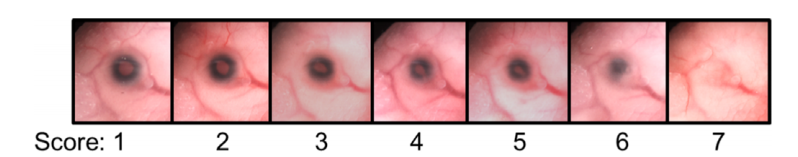
The eye deficits were analyzed and graded on a scale from 1 to 7, which can be seen below.

Brain tissue was removed from the fetuses and assessed for hydrocephalus. Hydrocephalus is a build up of cerebrospinal fluid in the brain. Normally, this is stored in special areas called the ventricles and circulated, but in a case of hydrocephalus, it accumulates and puts pressure on the brain.
Overall, it was found that fetuses with a knockout of the MNS1 gene that codes for the protein, or a defect to the MNS1 gene were more prone to facial defects, eye defects and hydrocephalus compared to the controls. There were no significant differences between the PAE group and the controls in terms of length or weight.
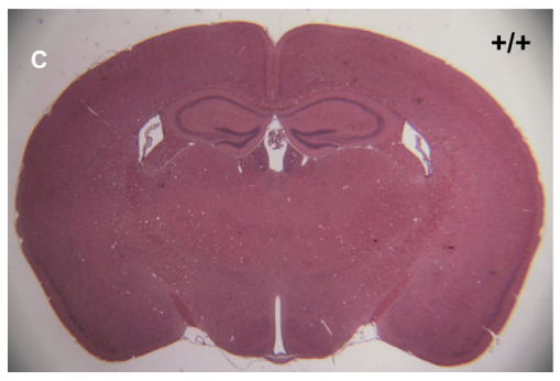
This is a sample of a normal brain from the mice at GD17. You can see the three ventricles (where the cerebrospinal fluid is stored) are normally sized, and the cortex (the pink area) has all the correct structures in the correct space. The cortex is responsible for controlling brain function and communication.
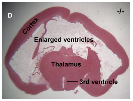
In this image, you can very see the expansion of the ventricles and the excess cerebrospinal fluid. The light space in the middle are the ventricles, which have merged together. You can also see an extreme reduction in the cortex, with little midbrain structures remaining.
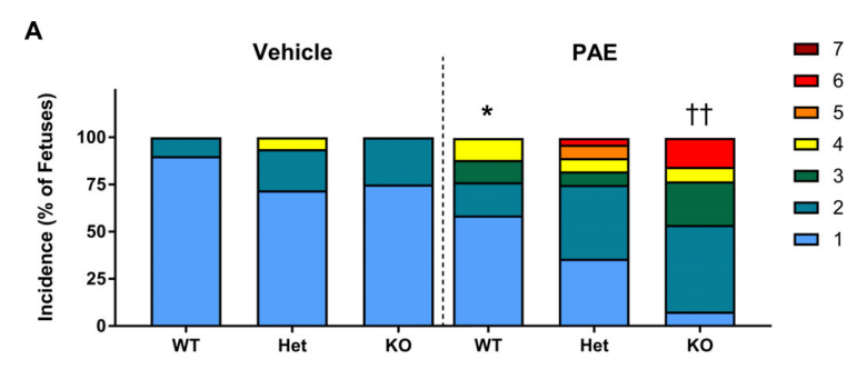
The above graph was generated by the authors and included in the paper. WT stands for wild type, which is a mouse that has no alterations made to the gene that codes for the MNS1 protein. Het stands for heterozygous. There are typically two copies of a gene that will code for a protein. With the heterozygous mice, they had one copy of the gene coding for the MNS1 protein altered, and one that was left completely normal. KO stands for knockout, these mice have had the gene coding for the MNS1 protein completely removed. The coloured scale on the right is the gradient of eye deformities, which is the same scale as the one showed before. As you can see in the graph, the mice with PAE have higher instances of more severe eye deficits compared to the control group.
In mice with eye deficits, there was also an increase in craniofacial deformities that can be correlated with the facial deformities seen in humans with FAS.
The results of this study suggest that the loss of the Mns1 gene increases the susceptibility of eye, craniofacial and central nervous system deformities.
The deformities may be caused in part by the disruption of proper motile cilia function. In normal development, cilia distribute morphogens (a substance that helps determine the pattern of tissue formation) to develop the left and right axes. In previous studies, mice with knock out MNS1 proteins have increased cases of situs inversus. Situs inversus is a condition where organs can develop in a mirrored position to what is typical.

The diagram shows a normal person on the left and a person with situs inversus on the right. The liver can be used as a landmark; in a normal patient it is on the left (remember the diagram is showing a person facing you, ie their right is your left) and in a person with situs inversus the liver is on the left.
This study is important because it is the first study done looking at the Mns1 gene and its association with the symptoms of FAS. Although it is done in a model organism, the results can potentially be applied to humans. Knowing this, new therapeutic technologies can be created and implemented that directly targets this pathway. Also, preventative measures can be taken in utero if a mother is drinking during pregnancy that may help to correctly form the motile cilia.
Article Reference
Boschen, K.E., H. Gong, L.B. Murdaugh & S.E. Parnell (2018). Knockdown of Mns1 Increases Susceptibility to Cranfiofacial-Defects Following Gastrulation Stage Alcohol Exposure in Mice. Alcoholism; Clinical and Experimental Research. 42(11):2136-2143.
